- Blog
12/05/2026
Cancer is one of the deadliest diseases of our time, with 19 to 20 million people diagnosed each year worldwide. Mortality remains high, although it varies greatly depending on the type of cancer. In 2020, approximately 10 million people died from the disease worldwide. Strategies for cancer detection, monitoring, and treatment are evolving rapidly and have helped reduce mortality for many types of cancer (Stribbling & Ryan, 2022).
Despite these advances, the figures highlight the importance of continuing research to improve treatments.
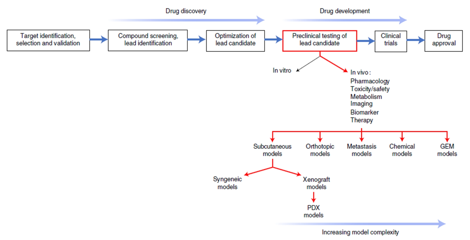
As with any drug development process, many steps are required before a therapeutic molecule can reach the market, and preclinical testing involving animals is one of them (see Figure 1).
Figure 1 : From Stribbling & Ryan, 2022. Preclinical cancer drug development. The steps involved in cancer drug development showing where cancer models fit into the testing of the lead candidate; rather than occurring sequentially, the steps involved in drug discovery and drug development often overlap. GEM, genetically engineered mouse.
In this context, mice are widely used for several reasons:
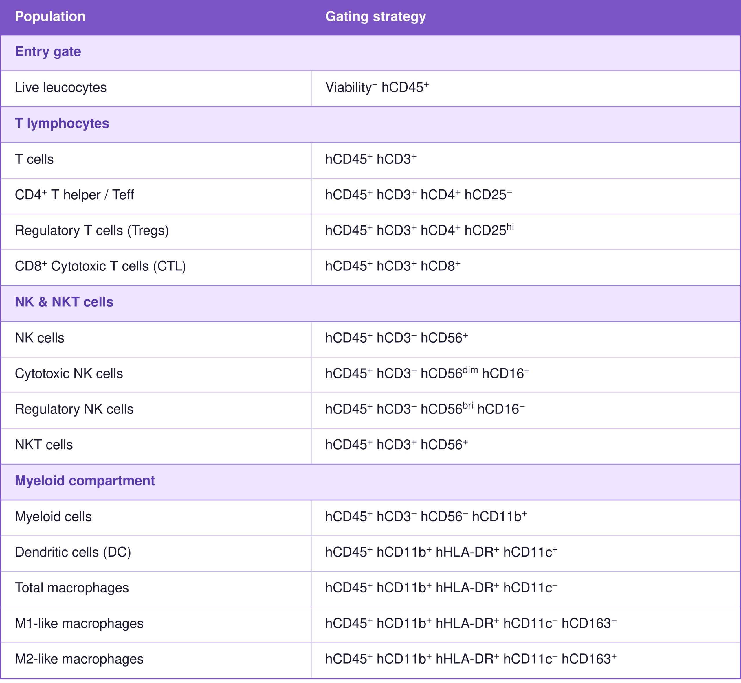
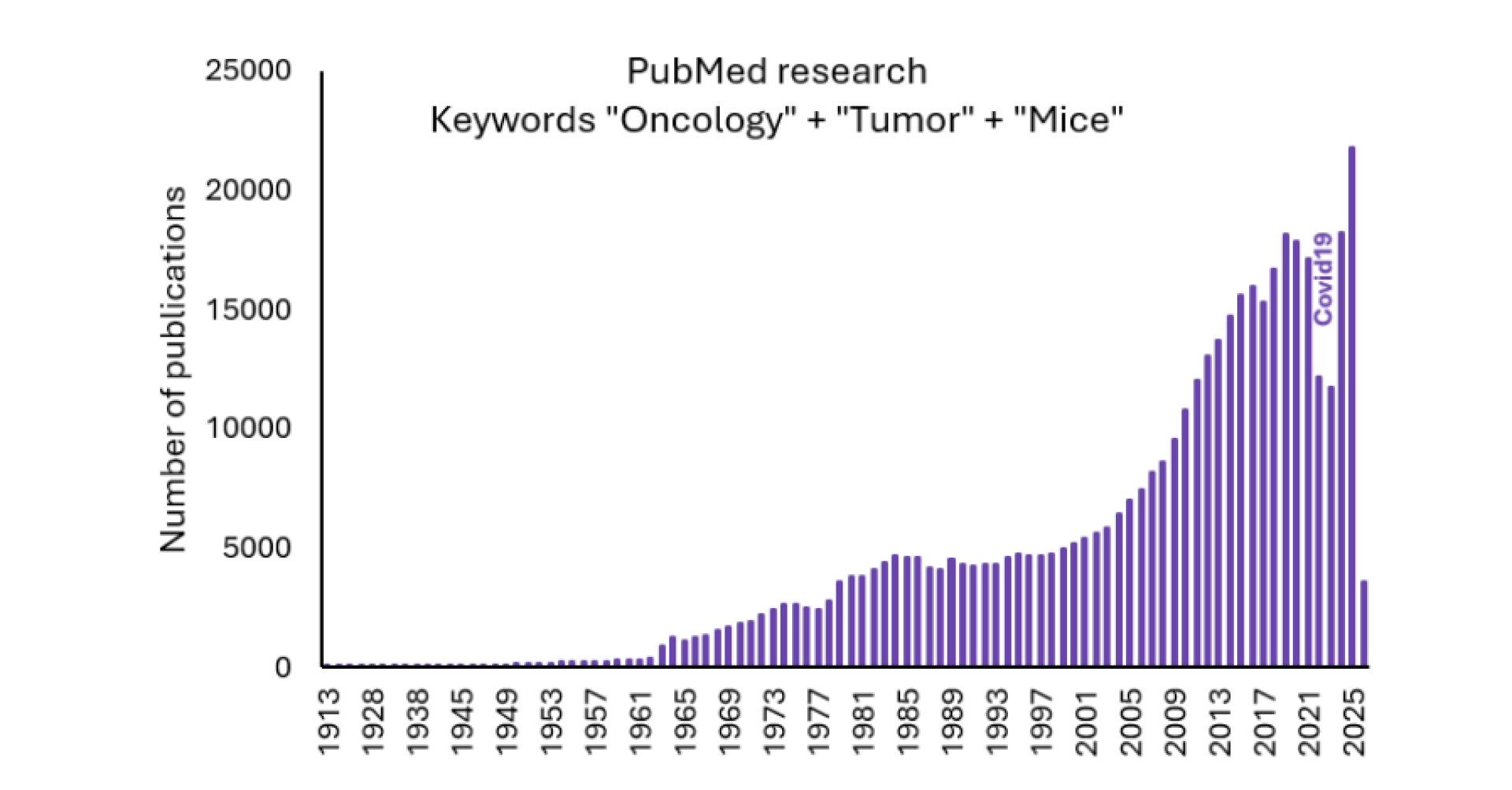
Immunodeficient and humanized mice (for example the models developed by TransCure bioServices) have been widely used in cancer research since the 1990s, with their use continuing to increase to this day (see Figure 2). These models make it possible to study interactions between tumors and the human immune system and to evaluate the potential of immunotherapies (De La Rochere et al., 2026).
Figure 2: Number of scientific publications containing the keywords “Oncology,” “Tumor,” and “Mice” over time.
There are many different types of cancer, so the methods used to study them are equally diverse.
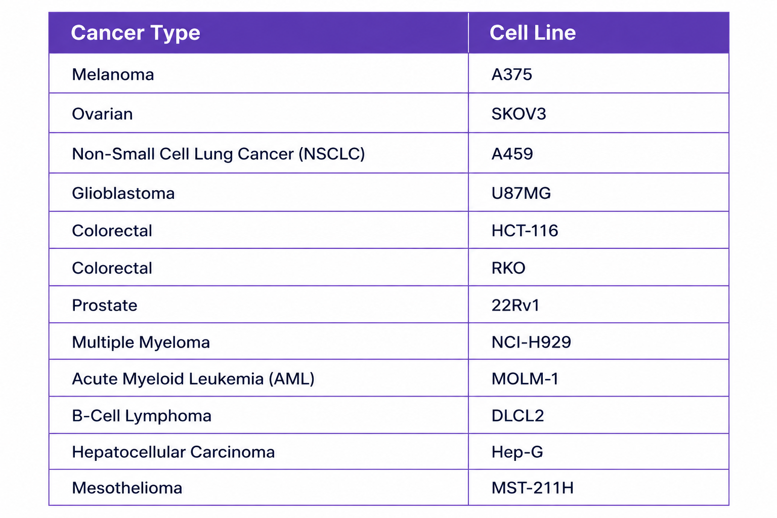
Tumors implanted in the flanks of mice are among the most commonly used models[1]. These tumors may originate from either patients or mice and can be implanted by injecting cells or surgically grafting a piece of tumor tissue (Cocco & de Stanchina, 2024).
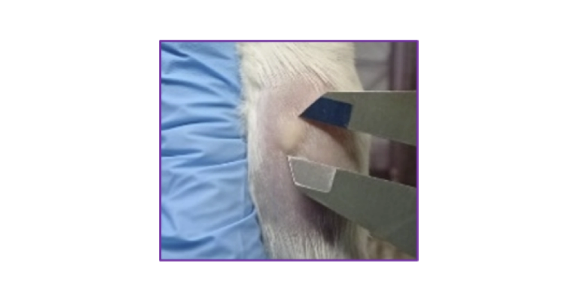
The subcutaneous tumor then grows and can easily be monitored using calipers to estimate its volume (see picture).
[1] To study a disease, we use a “model” of that condition. In other words, the causes or symptoms are reproduced in an animal. If the animal is a mouse, we refer to it as a murine model.
In some cases, these subcutaneous tumors may ulcerate*.
*Ulceration is defined as a pathological process involving tissue damage that results in the formation of an ulcer (a wound caused by tissue loss).
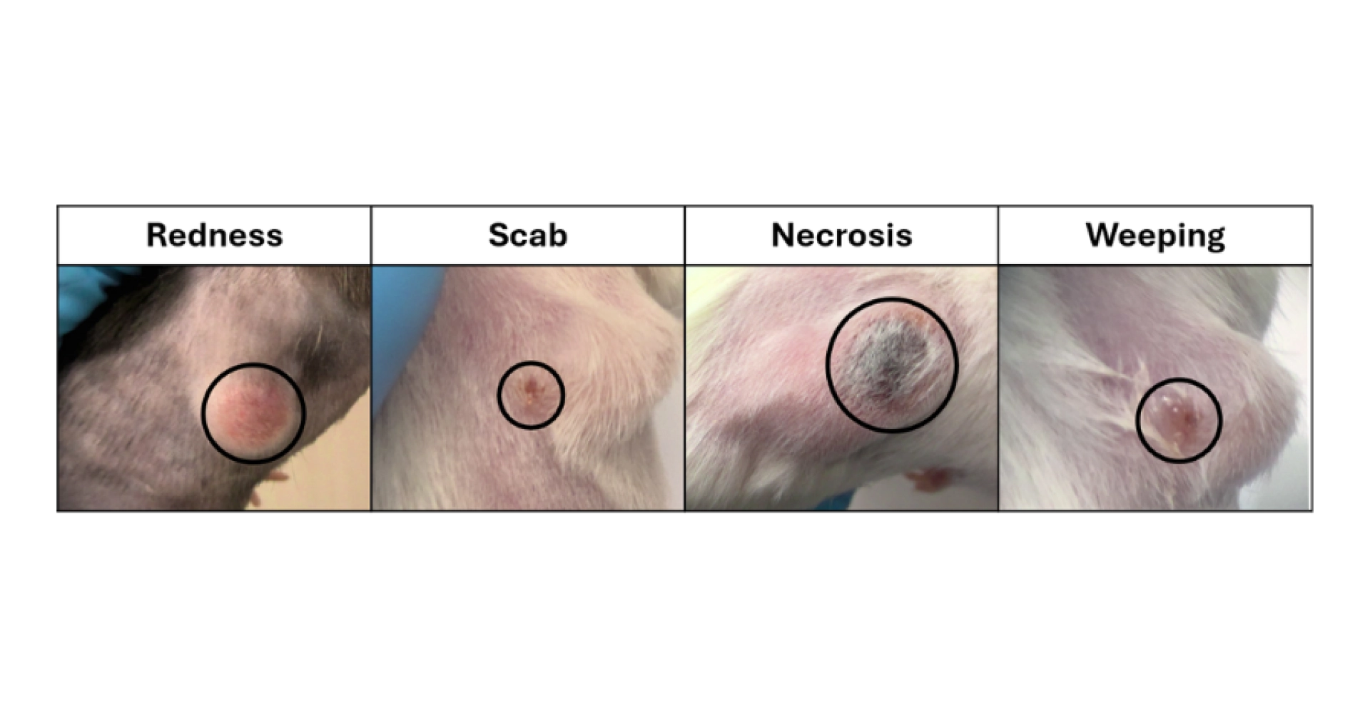
In the context of subcutaneous tumors, an ulcer corresponds to the loss of the epidermis and at least part of the dermis. Ulceration may be caused by, or associated with, necrosis (cell death) and/or inflammation of the surrounding tissues. The presence of a scab or a crater indicates that an ulcer is already established (UBC ACC, 2018). Various types of discharge may also be observed (see Figure 3).
Figure 3 : Examples of subcutaneous tumors that are ulcerated or in the process of ulcerating
All stages of ulceration may be associated with pain and should therefore be managed appropriately unless there is a scientifically justified reason not to do so (for example, in pain studies) (European parliament and of the council of 22 September 2010 on the protection of animals used for scientific purposes, 2010).
The factors contributing to tumor ulceration are numerous and still poorly understood (Stribbling & Ryan, 2022; UBC ACC, 2018):
The frequency of this adverse event varies greatly depending on these factors. In recent years, we have observed ulceration in approximately 10% of tumors across all stages combined.
Tumor ulceration is an important ethical concern. Animals exhibiting this clinical sign are considered to fall within the severe severity category.
At TransCure bioServices, addressing this issue is part of a broader commitment to animal welfare and the continuous improvement of experimental practices.
In France, oncology studies represent the fifth largest category of animal use classified as severe, and mice are the species most frequently affected by this severity category (see Table A4 in Annex 1 of the FC3R report on severe severity).
The same report shows that, across Europe, 5.6% of animal uses are related to oncology (Table A5), while 10.5% of severe procedures are oncology-related (Table A6) (FC3R Groupe de travail « Sévérité », 2026).
Although the causes of tumor ulceration remain poorly understood, several approaches can be implemented to:
1 – How can under occurrence be reduced?
Choice of cell line
As mentioned above, some cell lines ulcerate more frequently than others. In a CRO (Contract Research Organization) setting, many different cell lines may be used, with varying frequencies. Systematically recording ulcer occurrence makes it possible to identify the cell lines most at risk and therefore discourage their use with clients.
Quality control of cells before injection
Tumors generated from contaminated cells are more likely to ulcerate. Systematic testing of cells before injection eliminates this risk factor.
Cell injection technique
Needle type, bevel orientation, and technical details such as rotating the needle a quarter turn after injection before withdrawing it may all influence ulcer formation.
At TransCure bioServices, a modification of the injection procedure implemented in 2022 reduced the ulceration rate of flank subcutaneous tumors by approximately 50%.
2 – How can ulcer progression be slowed?
In general, a tumor that will eventually ulcerate first develops redness, a sign of tissue inflammation. A scab may then form. Depending on tumor growth rate and the discomfort experienced by the animal (leading to licking or scratching), the redness and/or scab may enlarge and develop into an increasingly severe lesion. The tumor may then develop a crater and become moist.
This stage is generally considered a humane endpoint (as is the case at TransCure bioServices), and the mouse must be euthanized.
In some cases, tissue death occurs and the tumor turns black. This is necrosis, which tends to spread over time.
Once a tumor begins to show redness, subsequent stages may develop more or less rapidly. Preventing this progression is difficult, especially if the tumor continues to grow.
Certain creams may help by promoting wound healing.
At TransCure bioServices, however, internal studies did not demonstrate any beneficial effect under our conditions of use.
Physical trauma may worsen lesions. Pain relief may help reduce this risk by decreasing discomfort and therefore reducing scratching behavior. This topic is discussed in the next section.
3 – How can pain be reduced?
Detecting pain in mice is challenging (see blog article “How to Effectively Assess the Well-being of Laboratory Mice”). According to the European Directive, suspected pain should be managed with appropriate analgesia (European parliament and of the council of 22 September 2010 on the protection of animals used for scientific purposes, 2010).
The different stages of ulceration (redness = inflammation, blackening = necrosis, weeping = potential infection) may all be painful.
When adjusted for body surface area, a 5 mm diameter lesion in a 30 g mouse would correspond to a lesion of more than 2 cm in a human (reference used: 1.60 m, 55 kg) (Cheung et al., 2009). It is difficult to imagine that a wound of this size would be painless in a mammal that possesses nociceptive systems similar to those of humans.
It is therefore essential to minimize pain in animals with tumor ulceration.
Because these lesions require medium-term management, the most appropriate approach is generally a systemic route of administration requiring as little intervention as possible.
At TransCure bioServices, buprenorphine is administered in the drinking water as soon as one of the signs of tumor ulceration appears (redness, scab formation, necrosis), unless a scientific contraindication has been demonstrated.
To ensure that lesions are managed as early as possible, animal care technicians must be trained to recognize them. Regular refresher training helps standardize practices and maintain optimal scientific quality.
Managing tumor ulceration requires a multifaceted approach.
As part of a continuous improvement strategy, TransCure bioServices has implemented several measures (see below):
It remains necessary to always strive to eliminate tumor ulceration, for the well-being of the animals and the researchers, and to ensure robust and reproducible scientific results.

Cheung, M. C., Spalding, P. B., Gutierrez, J. C., Balkan, W., Namias, N., Koniaris, L. G., & Zimmers, T. A. (2009). Body Surface Area Prediction in Normal, Hypermuscular, and Obese Mice. Journal of Surgical Research, 153(2), 326–331. https://doi.org/10.1016/J.JSS.2008.05.002
Cocco, E., & de Stanchina, E. (2024). Patient-Derived-Xenografts in Mice: A Preclinical Platform for Cancer Research. Cold Spring Harbor Perspectives in Medicine, 14(7). https://doi.org/10.1101/cshperspect.a041381
De La Rochere, P., Loumagne, L., Rathaux, M., Dubois, M., Denizeau, J., Nemati, F., Viel, S., Rocha, D., Slavnic, T., Thatte, J., Qixiang Li, H., Ouyang, X., Sedlik, C., Decaudin, D., Azar, G., Sidhu, S., Piaggio, E., Das, S., Yang, W., … La Rochere, D. P. (2026). A comprehensive analysis of humanized mouse models for the study of cancer immunotherapies. Frontiers in Immunology, 17, 1730378. https://doi.org/10.3389/FIMMU.2026.1730378
European parliament and of the council of 22 September 2010 on the protection of animals used for scientific purposes. (2010). Directive 2010/63/EU.
FC3R Groupe de travail « Sévérité ». (2026). Etat des lieux, comparaisons européennes, constats et recommandations. In Rapport du groupe de travail « Sévérité ». www.fc3r.com
Stribbling, S. M., & Ryan, A. J. (2022). The cell-line-derived subcutaneous tumor model in preclinical cancer research. Nature Protocols 2022 17:9, 17(9), 2108–2128. https://doi.org/10.1038/s41596-022-00709-3
UBC ACC. (2018). Guideline on Rodents with Ulcerated Subcutaneous Tumours: Protocol Requirements, Monitoring, Managing and Humane Endpoints. Animal Care Committee of the University of British Columbia.